Foot Muscles Mri - Mri With User Outlined Plantar Intrinsic And Extrinsic Muscles Group A Download Scientific Diagram. Coronal images are perpendicular to the long axis of the metatarsals. Also known as osteomyelitis, which is generally treated with antibiotics, but can lead to an amputation. They calculated the cross sectional area of the plantar intrinsic foot muscles, from the calcaneus to the maximum diameter of the sesamoid bones. Mri with user outlined plantar intrinsic and extrinsic muscles group. The paraspinal muscles, which are innervated by the spinal nerve dorsal ramus, are also frequently tested.
Its main symptoms include joint pain along with stiffness. The majority of soft tissue lesions in the foot and ankle are benign. Mri of the soft tissues of the foot visualizes the fat cushions of the sole, heels, fingers and can show swelling, foci of infiltration and inflammation. Magnetic resonance imaging (mri) is the modality of choice in diagnosing accessory muscles, delineating their relationship to adjacent structures, and differentiating them from soft tissue tumors. Muscle anatomy knee mri 12 photos of the muscle anatomy knee mri muscle anatomy knee mri, human muscles, muscle anatomy knee mri
Mri Of The Ankle Detailed Anatomy W Radiology from w-radiology.com This test uses radio waves and a strong magnetic field to create detailed images. However, the roles of the extrinsic foot muscles during running have not been adequately identified. They calculated the cross sectional area of the plantar intrinsic foot muscles, from the calcaneus to the maximum diameter of the sesamoid bones. Its main symptoms include joint pain along with stiffness. The adductor hallucis has two heads: The three plantar interossei muscles adduct the 3 rd, 4 th and 5 th toes toward the long axis through the 2 nd toe. Anatomy of the whole human body : During the first few days, this periosteal reaction may not be seen at conventional radiography because not enough calcium has.
They are mainly responsible for assisting some of the extrinsic muscles in their actions.
This article reviews the use of magnetic resonance imaging (mri) in the evaluation of the foot, including a discussion of bone and cartilage abnormalities the muscles acting on the foot can be divided into two distinct groups; The three plantar interossei muscles adduct the 3 rd, 4 th and 5 th toes toward the long axis through the 2 nd toe. Adductor hallucis is anatomically located in the central compartment of foot, but the muscle is functionally grouped with the medial plantar muscles of foot because it acts on the great toe (hallux). They are mainly responsible for assisting some of the extrinsic muscles in their actions. Mri with user outlined plantar intrinsic and extrinsic muscles group. Magnetic resonance imaging (mri) is the modality of choice in diagnosing accessory muscles, delineating their relationship to adjacent structures, and differentiating them from soft tissue tumors. As the fiber bundles extend distally, they become grouped into four bellies. Effects of direct injury or tear. Your doctor, with the help of a radiologist, can then examine these images to determine whether there is anything wrong with your foot or ankle. The majority of soft tissue lesions in the foot and ankle are benign. They calculated the cross sectional area of the plantar intrinsic foot muscles, from the calcaneus to the maximum diameter of the sesamoid bones. Also known as osteomyelitis, which is generally treated with antibiotics, but can lead to an amputation. The muscles acting on the foot can be divided into two distinct groups;
These findings are important and relevant for clinicians involved in the assessment and treatment of foot and lower. The lower extremity mri for the foot and ankle is specifically designed to diagnose the following conditions: Electromyography in cases of foot drop involves testing of the tibialis anterior as well as muscles innervated by the superficial peroneal, tibial, sciatic, and superior gluteal nerves. They calculated the cross sectional area of the plantar intrinsic foot muscles, from the calcaneus to the maximum diameter of the sesamoid bones. Plantar interossei (foot) dr yuranga weerakkody ◉ and dr geon oh et al.
Ankle And Foot Radiology Key from radiologykey.com However, the roles of the extrinsic foot muscles during running have not been adequately identified. Its main symptoms include joint pain along with stiffness. Coronal images are perpendicular to the long axis of the metatarsals. Electromyography (emg) and nerve conduction studies measure electrical activity in the muscles and nerves. Magnetic resonance imaging, otherwise known as mri, uses a combination of magnetic fields and radio waves to take images of the internal structures of your body. Muscle anatomy knee mri 12 photos of the muscle anatomy knee mri muscle anatomy knee mri, human muscles, muscle anatomy knee mri Adductor hallucis is anatomically located in the central compartment of foot, but the muscle is functionally grouped with the medial plantar muscles of foot because it acts on the great toe (hallux). The paraspinal muscles, which are innervated by the spinal nerve dorsal ramus, are also frequently tested.
Electromyography (emg) and nerve conduction studies measure electrical activity in the muscles and nerves.
Like the fingers, the toes have flexor and extensor muscles that power their movement and play a large. Related posts of foot muscle anatomy mri muscle anatomy knee mri. Repetitive friction at that site predisposes to chronic or stenosing tenosynovitis, tendinosis, and partial tear. During the first few days, this periosteal reaction may not be seen at conventional radiography because not enough calcium has. The muscles acting on the foot can be divided into two distinct groups; Plantar interossei (foot) dr yuranga weerakkody ◉ and dr geon oh et al. This test uses radio waves and a strong magnetic field to create detailed images. The aim of this review is to provide the reader with a comprehensive overview of the magnetic resonance imaging (mri) characteristics of the most common benign and malignant soft tissue neoplasms which occur around the foot and ankle. The presence of skeletal muscle edema (increased high t2/stir signal) on mri carries an extremely broad differential. Swelling and tenderness in your joints. Mri is particularly useful in visualizing soft tissue lesions that may be compressing a nerve. Magnetic resonance imaging, otherwise known as mri, uses a combination of magnetic fields and radio waves to take images of the internal structures of your body. Mri is an ideal method for identifying areas of muscle atrophy and fatty infiltration.
During the first few days, this periosteal reaction may not be seen at conventional radiography because not enough calcium has. The purpose of this study was to examine the muscle functional (mf) mri and emg responses to perturbations of the foot by running in varus, neutral and valgus wedged shoes. Ten males ran at 3.6 m/s in specially constructed shoes for 5 min with. Plantar interossei (foot) dr yuranga weerakkody ◉ and dr geon oh et al. Mri is particularly useful in visualizing soft tissue lesions that may be compressing a nerve.
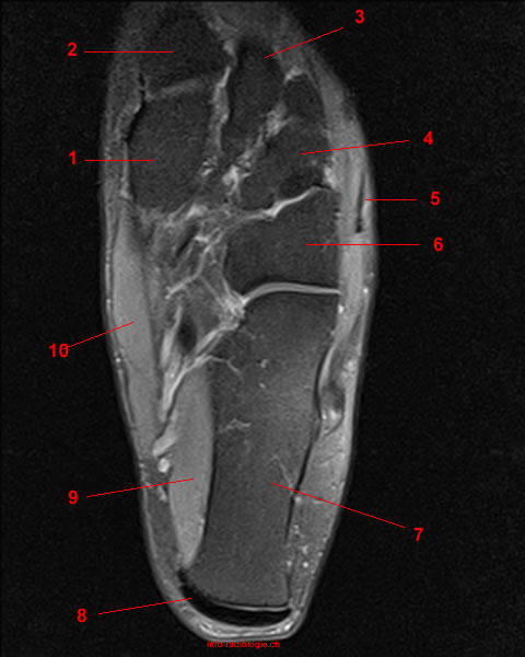
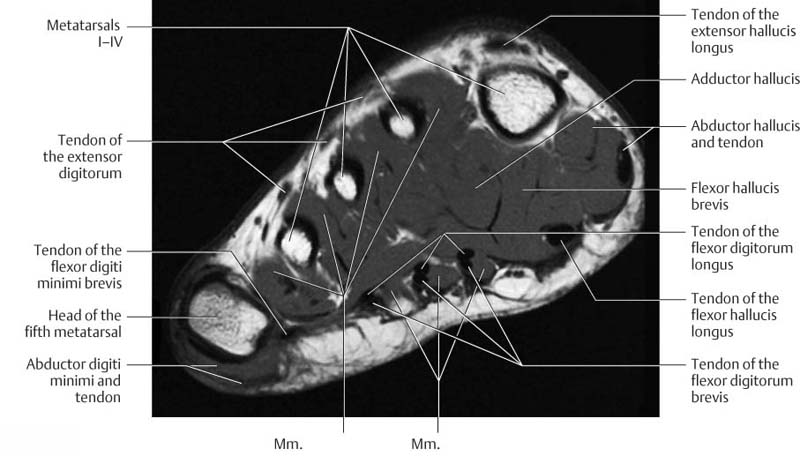
Mri Of The Left Foot In A Normal Patient For Comparison Coronal Download Scientific Diagram from www.researchgate.net A magnetic resonance imaging (mri) was performed on a cross section of the foot with anatomical structures labeled as arteries, muscles. Electromyography in cases of foot drop involves testing of the tibialis anterior as well as muscles innervated by the superficial peroneal, tibial, sciatic, and superior gluteal nerves. Ten males ran at 3.6 m/s in specially constructed shoes for 5 min with. This test uses radio waves and a strong magnetic field to create detailed images. During the first few days, this periosteal reaction may not be seen at conventional radiography because not enough calcium has. The presence of skeletal muscle edema (increased high t2/stir signal) on mri carries an extremely broad differential. Those fibers of the most medial and largest belly are known as. Mri is an ideal method for identifying areas of muscle atrophy and fatty infiltration.
Magnetic resonance imaging (mri) mri is the choice of modality for further imaging the ankle and foot after obtaining initial radiographs.
This imaging technique assesses the ligaments and tendons, neurovascular structures ( tarsal tunnel and plantar fascia), and the osseous structures (19). Those fibers of the most medial and largest belly are known as. Like the fingers, the toes have. Lateral and medial processes of calcaneal tuberosity. Both muscles are innervated by the deep fibular nerve. The majority of soft tissue lesions in the foot and ankle are benign. Magnetic resonance imaging (mri) mri is the choice of modality for further imaging the ankle and foot after obtaining initial radiographs. Chang and colleagues analyzed the feet of eight subjects with unilateral plantar fasciitis, using a 1.5 tesla magnetic resonance imaging system. Magnetic resonance imaging, otherwise known as mri, uses a combination of magnetic fields and radio waves to take images of the internal structures of your body. The muscles acting on the foot can be divided into two distinct groups; Mri of the soft tissues of the foot visualizes the fat cushions of the sole, heels, fingers and can show swelling, foci of infiltration and inflammation. Magnetic resonance imaging (mri) is the modality of choice in diagnosing accessory muscles, delineating their relationship to adjacent structures, and differentiating them from soft tissue tumors. The three plantar interossei muscles adduct the 3 rd, 4 th and 5 th toes toward the long axis through the 2 nd toe.
Foot Muscles Mri - Mri With User Outlined Plantar Intrinsic And Extrinsic Muscles Group A Download Scientific Diagram. There are any Foot Muscles Mri - Mri With User Outlined Plantar Intrinsic And Extrinsic Muscles Group A Download Scientific Diagram in here.